
Pap Smear using liquid-based cytology (LBC)
Pap tests are primarily performed to detect early signs of cervical cancer. However, most abnormal pap test results do not indicate cancer but show other underlying issues, such as a yeast infection, HPV infection, hormonal changes, or other issues. Dr Baloyi uses Liquid-based cytology (LBC), a new method of cervical cell sample preparation. The samples are collected in the usual way, but a brush-like device is used rather than a spatula. LBC based cervical cancer screening is generally more comfortable than a conventional smear based one.
Colposcopy and colposcopy guided biopsy
A colposcopy is an examination performed in the office in much the same way as a pelvic exam. During the procedure, a special magnifying device called a colposcope enables Dr Baloyi to clearly view abnormal areas of tissue on or around the cervix. During the examination, the patient lies on her back, and a lubricated speculum is inserted into the vagina. A solution may also be applied to the cervix and walls of the vaginal canal. Next, the colposcope is placed at the vaginal opening, and a bright light is shone onto the cervix and the vagina. Dr Baloyi looks through the magnifying lens to evaluate the cervix for any areas of abnormal tissue. If an abnormal area is identified, Dr Baloyi may take a small biopsy or tissue sample for further evaluation.
STD testing
STDs such as HIV, Syphilis and Hepatitis B can be diagnosed with a blood test. Vaginal swabs can test for Gonorrhoea, Chlamydia and Herpes and HPV. Diagnosis, management and treatment of STDs available for both you and your partner.
Endometrial screening with pipelle sampling
Endometrial pipelle sampling is a common procedure often performed in women with abnormal uterine bleeding. First, a special instrument called a speculum is inserted into the vagina. Next, A pipelle (a thin straw-like tube) is gently inserted through the cervix and the uterus. Dr Baloyi then gently moves the pipelle back and forth to obtain a sample. The procedure usually only takes a few minutes.
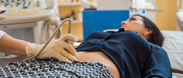
Ultrasounds
A gynaecological ultrasound is an assessment of the female pelvis, including the wall and lining of the uterus, the ovaries, and the endometrial lining.
There are two main types of pelvic ultrasounds; vaginal and abdominal.
During a vaginal ultrasound, a narrow, gel-covered probe is gently placed into the vagina. Most patients experience no pain or only some mild discomfort.
An abdominal ultrasound is done through your abdomen. During the ultrasound, you will lie on an exam table. Dr Baloyi will apply some gel onto the transducer. He will gently run the transducer back and forth over the skin of your belly.